An Ophthalmoscope Is Used to Determine Which of the Following
Damage to the optic nerve Retinal tear or detachment Glaucoma Macular degenerations Melanoma Diabetic retinopathy Hypertension Infection More advanced ophthalmoscopes offer doctors the ability to alter the aperture lens and aperturefilter combinations to gain a larger view of the fundus. Ophthalmoscopy also called fundoscopy or a fundoscopic exam is a common procedure performed by an eye doctor.
Moran Core How To Use The Direct Ophthalmoscope
Classic Migraine See also Chap.
. ADC Pocket Ophthalmoscopes have five aperture selections shown above. Have higher light intensity b. Following would determine refraction exam.
In an exam once you have found an abnormality keep looking for a second one. Reviewed by Brian Boxer Wachler MD. Which of the following is considered when preparing to examine an older adult.
This should be followed by a distant direct examination at 22-25cm a comfortable near vision distance. Which of the following is considered when preparing to examine an older adult. You will either lie.
An expected part of every eye exam ophthalmoscopes are. They are used all over the world and are an essential piece of apparatus for all who wish to study the intricate biology of the eye. The health care provider performs this exam by shining a beam of light through the pupil using an instrument called an ophthalmoscope.
The exam involves the use of special lenses and bright direct light to provide a better. An ophthalmoscope is about the size of a flashlight. All of the above B Halogen illumination does not a.
How would the medical assistant prepare the ophthalmoscope for use. Likewise the fundus is the only location where vasculature can be visualized. Regardless of model type these hand-held devices are critical in the evaluation and diagnosis of a variety of diseases in the eye.
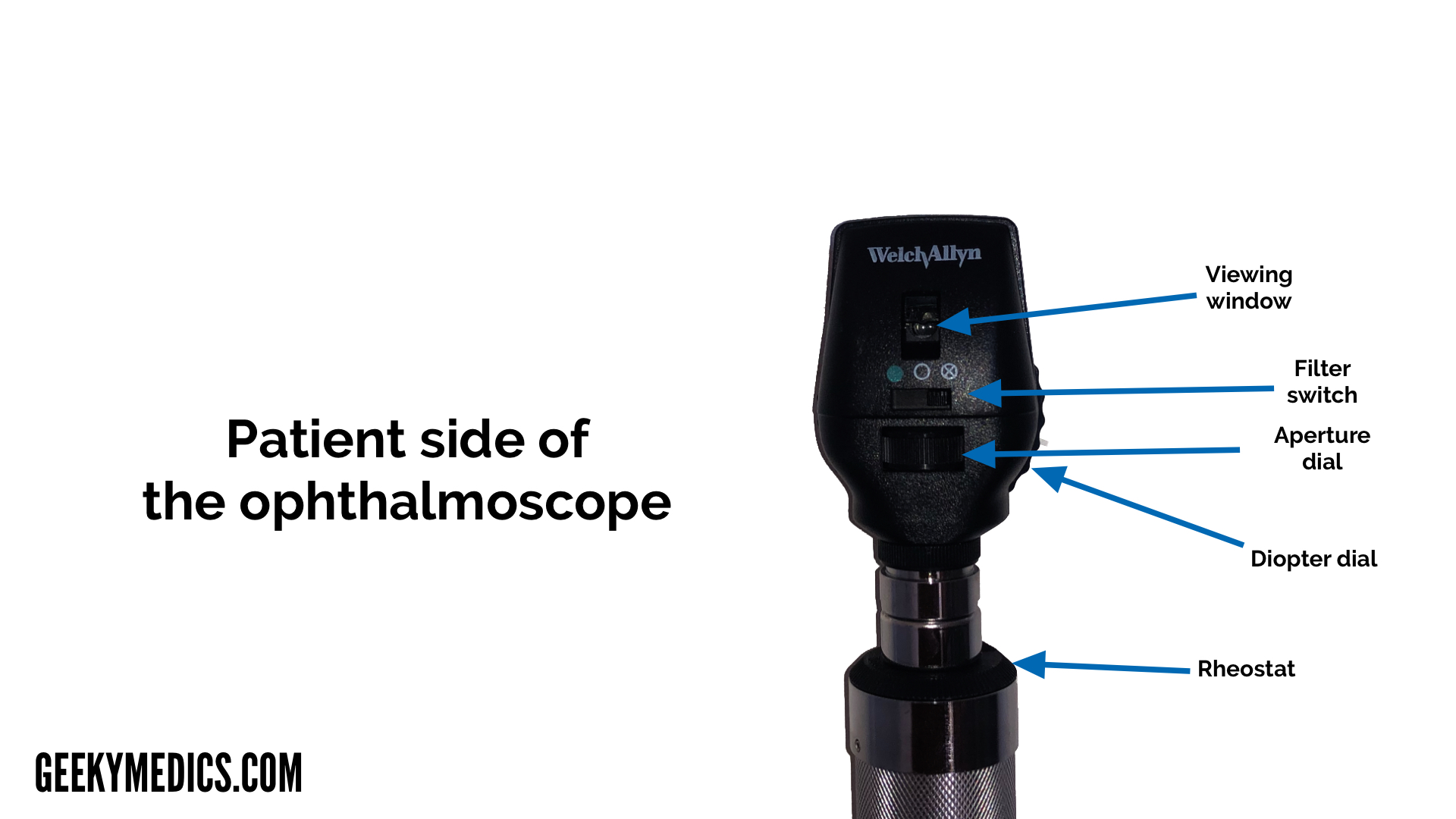
Provide more accurate representation of color A. Small spot large spot semicircle red-free filter and fixed. Improve communication with hearing-impaired.
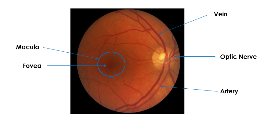
You will be seated in a darkened room. Red reflex anterior segment disc vessels and lastly macula see box. An ophthalmoscope is used for a funduscopic examination an examination of the internal c internal structures of the eye.
Ophthalmoscopy is done as part of a routine physical or complete eye examination mainly done by optometrists and ophthalmologists. Your eye doctor can look through the lenses to examine your eye. Traditionally part of almost every eye exam ophthalmoscopes can identify healthy structures within the eyeball and easily help your eye doctor see symptoms or indicators of diseases of the eye.
ABase the pace of. Ophthalmoscope pictures fundus photographs and ultrasonographic findings supplemented by a fundus diagram provide the foundation for determining tumor size verifying tumor location and surrounding critical structures. ABase the pace of the examination on the patients needs and abilities.
The diagnosis is confirmed by ophthalmoscopic examination of the dilated eye. Proper use of an ophthalmoscope requires a bit of practice and familiarity with the functions of your device. Avoid physical touch to offset making the older adult uncomfortable.
Introduction to the Fundoscopic Ophthalmoscopic Exam. Verify doubtful papillary action c. So much of what we see in internal medicine is vascular related and so viewing the fundus is a great way to get a sense for.
Base the pace of the examination on the patients needs and abilities. Detect lens opacities d. The retina is the only portion of the central nervous system visible from the exterior.
The first step in the use of an ophthalmoscope is to do examination at 1m distance. Touching the patients skin with the dorsal side of the hands and fingers. An ophthalmoscope is an instrument that has a light and several small lenses on it.
Mirror window Viewing aperture. It is used to detect and evaluate symptoms of various retinal vascular diseases or eye diseases such as glaucoma. 447 This usually occurs with a visual aura lasting about 20 min.
This sheds light on any abnormalities of the eyelids orbit and periorbita as well as highlights any obvious ocular deviations. Ophthalmoscopy also called fundoscopy is an exam your doctor optometrist or ophthalmologist uses to look into the back of your eye. Pediatricians and general practitioners may also include ophthalmoscopy in routine physical exams.
Which assessment technique should the nurse use to determine the body temperature of a patient. Have a longer useful life d. An ophthalmoscope is particularly useful for examining the structures of the retinathe light sensitive area at the back of the eye responsible for processing images.
Darken with use c. The ophthalmoscope can be used to. Ensure the batteries are charged _____ is the term used for a drooping of the upper eyelid.
An ophthalmoscope is a piece of equipment utilised by ophthalmologists that are used to inspect the internal structure of your eyes containing the retina. Detect foreign bodies in the cornea b. Which of the following is considered when preparing to examine an older adult.
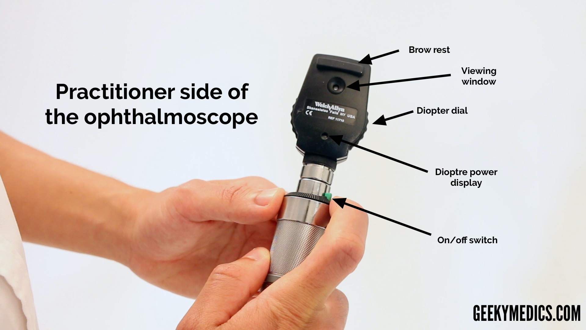
In a typical attack a small central disturbance in the field of vision marches toward the periphery leaving a transient scotoma in its wake. Which parts of the ophthalmoscope are present on the front end of the ophthalmoscope head. Structures of the eye.
Speak in clear low pitched tones. The ophthalmoscope can also be used for examining the anterior part of the eye by turning the lens dial to 10. A preliminary evaluation of adjacent critical structures can.
An ophthalmoscope can be used to check for. An ophthalmoscope is used for a funduscopic examination an examination of the internal structures of the eye. With it they can see the retina which senses light and.
What am I looking for. They may ask you to look in certain directions. An ophthalmoscope is used for a funduscopic examination an examination of the internal structures of the eye.
It has a light and different tiny lenses that allow the provider to view the back of the eyeball.
Fundoscopy Ophthalmoscopy Osce Guide Geeky Medics
Fundoscopy Ophthalmoscopy Osce Guide Geeky Medics

Moran Core How To Use The Direct Ophthalmoscope


Comments
Post a Comment